7D7O

Crystal structure of cystathionine gamma-lyase from Bacillus cereus ATCC 14579


Experimental Data Snapshot

  • Method: X-RAY DIFFRACTION
  • Resolution: 1.98 Å
  • R-Value Free: 0.262 
  • R-Value Work: 0.212 
  • R-Value Observed: 0.215 

wwPDB Validation   3D Report Full Report


Ligand Structure Quality Assessment 


This is version 1.1 of the entry. See complete history


Literature

Structural and Functional Characterization of Cystathionine gamma-lyase from Bacillus cereus ATCC 14579.

Sagong, H.Y.Kim, B.Joo, S.Kim, K.J.

(2020) J Agric Food Chem 68: 15267-15274

  • DOI: https://doi.org/10.1021/acs.jafc.0c06503
  • Primary Citation of Related Structures:  
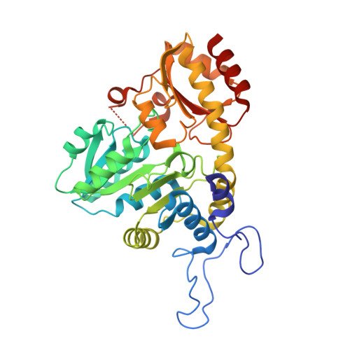
    7D7O

  • PubMed Abstract: 

    Cysteine is a semiessential amino acid and plays an important role in metabolism and protein structure and has also been applied in various industrial fields, such as pharmaceutical, food, cosmetic, and animal feed industries. Metabolic engineering studies have been conducted for the cysteine production through bacterial fermentation, but studies on the cysteine biosynthetic pathway in microorganisms are limited. We report the biochemical characteristics of cystathionine γ-lyase from Bacillus cereus ATCC 14579 ( Bc CGL). We also determined the crystal structure of Bc CGL in complex with the PLP cofactor and identified the substrate binding mode. We observed that the replacement of the conserved Glu321 residue to alanine showed increased activity by providing wider active site entrance and hydrophobic interaction for the substrate. We suggest that the structural differences of the α13-α14 region in CGL enzymes might determine the active site conformation.


  • Organizational Affiliation

    School of Life Sciences, KNU Creative BioResearch Group, Kyungpook National University, Daegu 41566, Republic of Korea.


Macromolecules
Find similar proteins by:  (by identity cutoff)  |  3D Structure
Entity ID: 1
MoleculeChains Sequence LengthOrganismDetailsImage
Bifunctional cystathionine gamma-lyase/homocysteine desulfhydrase
A, B
377Bacillus cereus ATCC 14579Mutation(s): 0 
Gene Names: EJ379_22345
UniProt
Find proteins for Q818A3 (Bacillus cereus (strain ATCC 14579 / DSM 31 / CCUG 7414 / JCM 2152 / NBRC 15305 / NCIMB 9373 / NCTC 2599 / NRRL B-3711))
Explore Q818A3 
Go to UniProtKB:  Q818A3
Entity Groups  
Sequence Clusters30% Identity50% Identity70% Identity90% Identity95% Identity100% Identity
UniProt GroupQ818A3
Sequence Annotations
Expand
  • Reference Sequence
Small Molecules
Ligands 3 Unique
IDChains Name / Formula / InChI Key2D Diagram3D Interactions
PLP (Subject of Investigation/LOI)
Query on PLP

Download Ideal Coordinates CCD File 
C [auth A],
I [auth B]
PYRIDOXAL-5'-PHOSPHATE
C8 H10 N O6 P
NGVDGCNFYWLIFO-UHFFFAOYSA-N
SO4
Query on SO4

Download Ideal Coordinates CCD File 
G [auth A],
H [auth A],
L [auth B],
M [auth B]
SULFATE ION
O4 S
QAOWNCQODCNURD-UHFFFAOYSA-L
GOL
Query on GOL

Download Ideal Coordinates CCD File 
D [auth A],
E [auth A],
F [auth A],
J [auth B],
K [auth B]
GLYCEROL
C3 H8 O3
PEDCQBHIVMGVHV-UHFFFAOYSA-N
Experimental Data & Validation

Experimental Data

  • Method: X-RAY DIFFRACTION
  • Resolution: 1.98 Å
  • R-Value Free: 0.262 
  • R-Value Work: 0.212 
  • R-Value Observed: 0.215 
  • Space Group: P 43 21 2
Unit Cell:
Length ( Å )Angle ( ˚ )
a = 116.708α = 90
b = 116.708β = 90
c = 147.794γ = 90
Software Package:
Software NamePurpose
HKL-2000data reduction
REFMACrefinement
PDB_EXTRACTdata extraction
HKL-2000data scaling
MOLREPphasing

Structure Validation

View Full Validation Report


Ligand Structure Quality Assessment 


Entry History & Funding Information

Deposition Data


Funding OrganizationLocationGrant Number
Rural Development AdministrationKorea, Republic OfPJ01492602

Revision History  (Full details and data files)

  • Version 1.0: 2021-08-18
    Type: Initial release
  • Version 1.1: 2023-11-29
    Changes: Data collection, Refinement description