Dual alpha-1,4- and beta-1,4-Glycosidase Activities by the Novel Carbohydrate-Binding Module in alpha-l-Fucosidase from Vibrio sp. Strain EJY3.
Hong, H., Kim, D.H., Seo, H., Kim, K.H., Kim, K.J.(2021) J Agric Food Chem 69: 3380-3389
- PubMed: 33705122
- DOI: https://doi.org/10.1021/acs.jafc.0c08199
- Primary Citation of Related Structures:
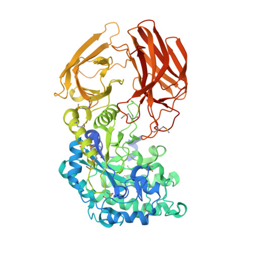
7DB5 - PubMed Abstract:
Carbohydrates are structurally and functionally diverse materials including polysaccharides, and marine organisms are known to have many enzymes for the breakdown of complex polysaccharides. Here, we identified an α-l-fucosidase enzyme from the marine bacterium Vibrio sp. strain EJY3 ( Vej FCD) that has dual α-1,4-glucosidic and β-1,4-galactosidic specificities. We determined the crystal structure of Vej FCD and provided the structural basis underlying the dual α- and β-glycosidase activities of the enzyme. Unlike other three-domain FCDs, in Vej FCD, carbohydrate-binding module-B (CBM-B) with a novel β-sandwich fold tightly contacts with the CatD/CBM-B main body and provides key residues for the β-1,4-glycosidase activity of the enzyme. The phylogenetic tree analysis suggests that only a few FCDs from marine microorganisms have the key structural features for dual α-1,4- and β-1,4-glycosidase activities. This study provides the structural insights into the mechanism underlying the novel glycoside hydrolase activities and could be applied for more efficient utilization in the hydrolysis of complex carbohydrates in biotechnological applications.
Organizational Affiliation:
School of Life Sciences, KNU Creative BioResearch Group, Kyungpook National University, Daehak-ro 80, Buk-ku, Daegu 41566, Republic of Korea.