6A6Q

Crystal structure of a lignin peroxidase isozyme H8 variant that is stable at very acidic pH


Experimental Data Snapshot

  • Method: X-RAY DIFFRACTION
  • Resolution: 1.67 Å
  • R-Value Free: 0.171 
  • R-Value Work: 0.141 
  • R-Value Observed: 0.143 

wwPDB Validation   3D Report Full Report


Ligand Structure Quality Assessment 


This is version 1.2 of the entry. See complete history


Literature

In silico-designed lignin peroxidase fromPhanerochaete chrysosporiumshows enhanced acid stability for depolymerization of lignin.

Pham, L.T.M.Seo, H.Kim, K.J.Kim, Y.H.

(2018) Biotechnol Biofuels 11: 325-325

  • DOI: https://doi.org/10.1186/s13068-018-1324-4
  • Primary Citation of Related Structures:  
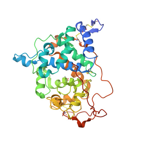
    6A6Q

  • PubMed Abstract: 

    The lignin peroxidase isozyme H8 from the white-rot fungus Phanerochaete chrysosporium (LiPH8) demonstrates a high redox potential and can efficiently catalyze the oxidation of veratryl alcohol, as well as the degradation of recalcitrant lignin. However, native LiPH8 is unstable under acidic pH conditions. This characteristic is a barrier to lignin depolymerization, as repolymerization of phenolic products occurs simultaneously at neutral pH. Because repolymerization of phenolics is repressed at acidic pH, a highly acid-stable LiPH8 could accelerate the selective depolymerization of recalcitrant lignin.


  • Organizational Affiliation

    1School of Energy and Chemical Engineering, UNIST, 50 UNIST-gil, Ulju-gun, Ulsan, 44919 Republic of Korea.


Macromolecules
Find similar proteins by:  (by identity cutoff)  |  3D Structure
Entity ID: 1
MoleculeChains Sequence LengthOrganismDetailsImage
Ligninase H8351Phanerochaete chrysosporium RP-78Mutation(s): 3 
Gene Names: LPOA
EC: 1.11.1.14
UniProt
Find proteins for P06181 (Phanerodontia chrysosporium)
Explore P06181 
Go to UniProtKB:  P06181
Entity Groups  
Sequence Clusters30% Identity50% Identity70% Identity90% Identity95% Identity100% Identity
UniProt GroupP06181
Sequence Annotations
Expand
  • Reference Sequence
Experimental Data & Validation

Experimental Data

  • Method: X-RAY DIFFRACTION
  • Resolution: 1.67 Å
  • R-Value Free: 0.171 
  • R-Value Work: 0.141 
  • R-Value Observed: 0.143 
  • Space Group: P 1 21 1
Unit Cell:
Length ( Å )Angle ( ˚ )
a = 41.212α = 90
b = 99.621β = 113.86
c = 48.322γ = 90
Software Package:
Software NamePurpose
REFMACrefinement
PDB_EXTRACTdata extraction
HKL-2000data reduction
HKL-2000data scaling
MOLREPphasing

Structure Validation

View Full Validation Report


Ligand Structure Quality Assessment 


Entry History 

Deposition Data

Revision History  (Full details and data files)

  • Version 1.0: 2019-01-23
    Type: Initial release
  • Version 1.1: 2020-09-16
    Changes: Derived calculations, Structure summary
  • Version 1.2: 2023-11-22
    Changes: Data collection, Database references, Refinement description