5E3Q

Crystal structure of DapD in complex with succinyl-CoA from Corynebacterium glutamicum


Experimental Data Snapshot

  • Method: X-RAY DIFFRACTION
  • Resolution: 1.80 Å
  • R-Value Free: 0.187 
  • R-Value Work: 0.157 
  • R-Value Observed: 0.158 

wwPDB Validation   3D Report Full Report


Ligand Structure Quality Assessment 


This is version 1.3 of the entry. See complete history


Literature

Crystal Structure and Biochemical Characterization of Tetrahydrodipicolinate N-Succinyltransferase from Corynebacterium glutamicum.

Sagong, H.Y.Kim, K.J.

(2015) J Agric Food Chem 63: 10641-10646

  • DOI: https://doi.org/10.1021/acs.jafc.5b04785
  • Primary Citation of Related Structures:  
    5E3P, 5E3Q, 5E3R

  • PubMed Abstract: 

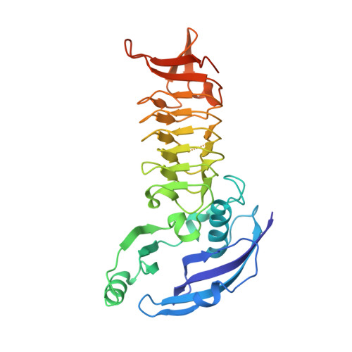
    Tetrahydrodipicolinate N-succinyltransferase (DapD) is an enzyme involved in the biosynthesis of l-lysine by converting tetrahydrodipicolinate into N-succinyl-l-2-amino-6-oxopimelate, using succinyl-CoA as a cofactor. We determined the crystal structure of DapD from Corynebacterium glutamicum (CgDapD). CgDapD functions as a trimer, and each monomer consists of three domains: an N-terminal helical domain (NTD), a left-handed β-helix (LβH) domain, and a β C-terminal domain (CTD). The mode of cofactor binding to CgDapD, elucidated by determining the structure in complex with succinyl-CoA, reveals that the position of the CTD changes slightly as the cofactor binds to the enzyme. The superposition of this structure with that of Mycobacterium tuberculosis shows differences in residues that make up cofactor-binding sites. Moreover, we determined the structure of CgDapD in complex with the substrate analogue 2-aminopimelate and revealed that the analogue was stabilized by conserved residues. The catalytic and substrate binding sites of CgDapD were confirmed by site-directed mutagenesis experiments.


  • Organizational Affiliation

    School of Life Sciences, KNU Creative BioResearch Group, Kyungpook National University , Daehak-ro 80, Buk-ku, Daegu 702-701, Korea.


Macromolecules
Find similar proteins by:  (by identity cutoff)  |  3D Structure
Entity ID: 1
MoleculeChains Sequence LengthOrganismDetailsImage
2,3,4,5-tetrahydropyridine-2,6-dicarboxylate N-succinyltransferase302Corynebacterium glutamicum ATCC 13032Mutation(s): 0 
Gene Names: dapDCgl1106cg1256
EC: 2.3.1.117
UniProt
Find proteins for Q8NRE3 (Corynebacterium glutamicum (strain ATCC 13032 / DSM 20300 / BCRC 11384 / JCM 1318 / LMG 3730 / NCIMB 10025))
Explore Q8NRE3 
Go to UniProtKB:  Q8NRE3
Entity Groups  
Sequence Clusters30% Identity50% Identity70% Identity90% Identity95% Identity100% Identity
UniProt GroupQ8NRE3
Sequence Annotations
Expand
  • Reference Sequence
Small Molecules
Ligands 1 Unique
IDChains Name / Formula / InChI Key2D Diagram3D Interactions
SCA
Query on SCA

Download Ideal Coordinates CCD File 
B [auth A]SUCCINYL-COENZYME A
C25 H40 N7 O19 P3 S
VNOYUJKHFWYWIR-ITIYDSSPSA-N
Experimental Data & Validation

Experimental Data

  • Method: X-RAY DIFFRACTION
  • Resolution: 1.80 Å
  • R-Value Free: 0.187 
  • R-Value Work: 0.157 
  • R-Value Observed: 0.158 
  • Space Group: H 3 2
Unit Cell:
Length ( Å )Angle ( ˚ )
a = 92.457α = 90
b = 92.457β = 90
c = 282.341γ = 120
Software Package:
Software NamePurpose
HKL-2000data collection
REFMACrefinement
PDB_EXTRACTdata extraction
HKL-2000data reduction
HKL-2000data scaling
CNSphasing

Structure Validation

View Full Validation Report


Ligand Structure Quality Assessment 


Entry History 

Deposition Data

Revision History  (Full details and data files)

  • Version 1.0: 2015-11-04
    Type: Initial release
  • Version 1.1: 2015-12-30
    Changes: Database references
  • Version 1.2: 2020-09-09
    Changes: Derived calculations, Structure summary
  • Version 1.3: 2023-11-08
    Changes: Data collection, Database references, Refinement description