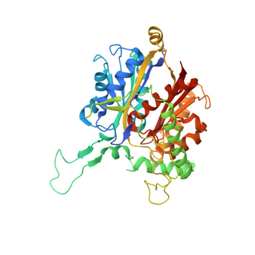
Crystal structure and biochemical characterization of beta-keto thiolase B from polyhydroxyalkanoate-producing bacterium Ralstonia eutropha H16
Kim, E.J., Son, H.F., Kim, S., Ahn, J.W., Kim, K.J.(2014) Biochem Biophys Res Commun 444: 365-369
- PubMed: 24462871
- DOI: https://doi.org/10.1016/j.bbrc.2014.01.055
- Primary Citation of Related Structures:
4NZS - PubMed Abstract:
ReBktB is a β-keto thiolase from Ralstonia eutropha H16 that catalyzes condensation reactions between acetyl-CoA with acyl-CoA molecules that contains different numbers of carbon atoms, such as acetyl-CoA, propionyl-CoA, and butyryl-CoA, to produce valuable bioproducts, such as polyhydroxybutyrate, polyhydroxybutyrate-hydroxyvalerate, and hexanoate. We solved a crystal structure of ReBktB at 2.3Å, and the overall structure has a similar fold to that of type II biosynthetic thiolases, such as PhbA from Zoogloea ramigera (ZrPhbA). The superposition of this structure with that of ZrPhbA complexed with CoA revealed the residues that comprise the catalytic and substrate binding sites of ReBktB. The catalytic site of ReBktB contains three conserved residues, Cys90, His350, and Cys380, which may function as a covalent nucleophile, a general base, and second nucleophile, respectively. For substrate binding, ReBktB stabilized the ADP moiety of CoA in a distinct way compared to ZrPhbA with His219, Arg221, and Asp228 residues, whereas the stabilization of β-mercaptoethyamine and pantothenic acid moieties of CoA was quite similar between these two enzymes. Kinetic study of ReBktB revealed that K(m), V(max), and K(cat) values of 11.58 μM, 1.5 μmol/min, and 102.18 s(-1), respectively, and the catalytic and substrate binding sites of ReBktB were further confirmed by site-directed mutagenesis experiments.
Organizational Affiliation:
Structural and Molecular Biology Laboratory, School of Life Sciences and Biotechnology, Kyungpook National University, Daehak-ro 80, Buk-ku, Daegu 702-701, Republic of Korea.


