


Crystal structure of Homoserine O-succinyltransferase from Escherichia coli K-12
Sagong, H.-Y., Kim, K.-J.To be published.
Experimental Data Snapshot
wwPDB Validation 3D Report Full Report

Entity ID: 1 | |||||
|---|---|---|---|---|---|
| Molecule | Chains | Sequence Length | Organism | Details | Image |
| Homoserine O-succinyltransferase | 309 | Escherichia coli K-12 | Mutation(s): 0 Gene Names: metAS, metA, b4013, JW3973 EC: 2.3.1.46 | 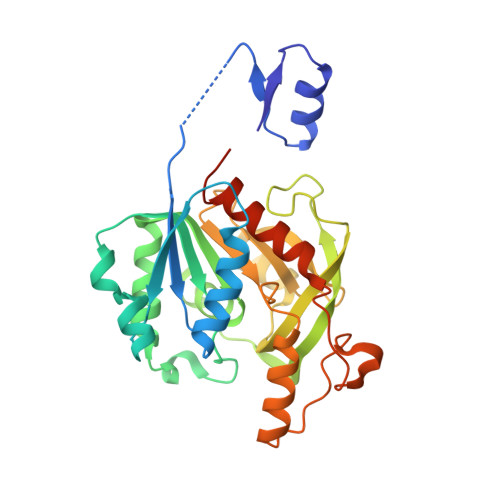 | |
UniProt | |||||
Find proteins for P07623 (Escherichia coli (strain K12)) Explore P07623 Go to UniProtKB: P07623 | |||||
Entity Groups | |||||
| Sequence Clusters | 30% Identity50% Identity70% Identity90% Identity95% Identity100% Identity | ||||
| UniProt Group | P07623 | ||||
Sequence AnnotationsExpand | |||||
| |||||
| Ligands 3 Unique | |||||
|---|---|---|---|---|---|
| ID | Chains | Name / Formula / InChI Key | 2D Diagram | 3D Interactions | |
| SO4 Query on SO4 | C [auth A], F [auth B] | SULFATE ION O4 S QAOWNCQODCNURD-UHFFFAOYSA-L |  | ||
| GOL Query on GOL | E [auth B] | GLYCEROL C3 H8 O3 PEDCQBHIVMGVHV-UHFFFAOYSA-N |  | ||
| EDO Query on EDO | D [auth A] | 1,2-ETHANEDIOL C2 H6 O2 LYCAIKOWRPUZTN-UHFFFAOYSA-N |  | ||
| Length ( Å ) | Angle ( ˚ ) |
|---|---|
| a = 76.348 | α = 90 |
| b = 81.027 | β = 90 |
| c = 98.093 | γ = 90 |
| Software Name | Purpose |
|---|---|
| REFMAC | refinement |
| HKL-2000 | data scaling |
| PDB_EXTRACT | data extraction |
| MOLREP | phasing |
| HKL-2000 | data reduction |
RCSB PDB (citation) is hosted by
RCSB PDB is a member of the

